Wikisage, the free encyclopedia of the second generation, is digital heritage
File:Illustrative samples of EEG from 14 children with Panayiotopoulos syndrome.jpg

Original file (440 × 667 pixels, file size: 163 KB, MIME type: image/jpeg)
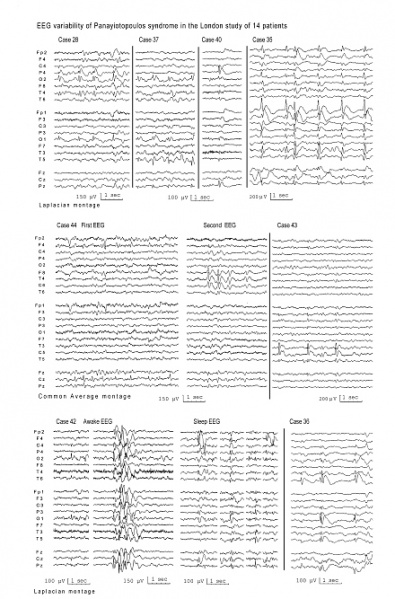
Illustrative samples of EEG from 14 children with Panayiotopoulos syndrome. Note that spikes may be localized in any and usually multiple brain regions (multifocal spikes). Occipital spikes are common (cases 28 and 37) but these are not a prerequisite for diagnosis (cases 40, 43, 44). Cloned-like repetitive multifocal spike-wave complexes, repetitive spike or sharp and slow wave complexes that appear concurrently in different brain locations of one or both hemispheres, may be abundant (case 35). Spike location may be shifting in serial EEGs (case 44). Brief generalized discharges may occur alone or with focal spikes (case 42).
File history
Click on a date/time to view the file as it appeared at that time.
| Date/Time | Thumbnail | Dimensions | User | Comment | |
|---|---|---|---|---|---|
| current | 01:22, 18 October 2015 | | 440 × 667 (163 KB) | Penarc (talk | contribs) | Illustrative samples of EEG from 14 children with Panayiotopoulos syndrome. Note that spikes may be localized in any and usually multiple brain regions (multifocal spikes). Occipital spikes are common (cases 28 and 37) but these are not a prerequisite... |
You cannot overwrite this file.
File usage
The following page uses this file: